Nikola Tesla Books
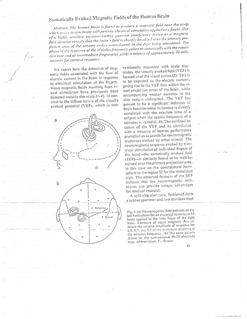
Somatically Evoked Magnetic Fields of the Human Brain Abstract. The human brain is found to produce a magnetic field near the sculp which varies in synchrony with periodic electrical stimulation applied to a finger. Use of a highly sensitive superconducting quantum interference device as a magnetic field detector reveals that the brain's field is sharply localized over the primary projection area of the sensory cortex contralateral to the digit being stimulated. The phase of the response at the stimulus frequency varies monotonically with the repetition rate and at intermediate frequencies yields a latency of approximately 70 milliseconds for cortical response. We report here the detection of magnetic fields associated with the flow of electric current in the brain in response to electrical stimulation of the fingers. Weak magnetic fields resulting from visual stimulation have previously been detected outside the scalp (14). In contrast to the diffuse nature of the visually evoked potential (VEP), which is cona F. Rolandic F. Sylvian ventionally measured with scalp electrodes, the visually evoked field (VEF) is located over the visual cortex (2). This is to be expected as the electric currents giving rise to the VEF flow within the visual projection areas of the brain, while accompanying weaker currents in the skin remain undetected. The VEF has proved to be a significant indicator of brain function since its latency is directly correlated with the reaction time of a subject when the spatial frequency of a stimulus is varied (5, 6). The confined location of the VEF and its correlation with a measure of human performance prompted us to search for neuromagnetic responses evoked by other stimuli. The neuromagnetic response evoked by electrical stimulation of individual fingers of the hand-the somatically evoked field (SEF) is similarly found to be well localized over the primary projection area. in this case on the contralateral hemisphere in the region SI for the stimulated digit. The observed features of the SEF indicate that the neuromagnetic techniques can provide unique advantages for medical research. A split-ring electrode, fashioned from a rubber grommet and two stainless steel Fig. 1. (a) Neuromagnetic field patterns on the left hemisphere for an electrical stimulus at 13 hertz applied to the little finger of the right hand. Contours of equal magnetic flux indicate the relative amplitude of response for 0.9.0.7, and 0.5 of the maximum response at the stimulus frequency. (b) The same pattern drawn on the conventional. 10-20 electrode map. Abbreviation: F., fissure. 81
